3D arm scanning with a mobile device now allows orthopedic professionals to accurately capture the morphology of the upper limb directly from a smartphone. It offers a fast, non-invasive, and highly efficient solution to design 100% custom medical devices. In the following guide, discover step by step how to perform a 3D arm scan using a mobile device.
Why perform a 3D arm scan with a mobile device?
A 3D scan of the upper limb allows for accurate digital modeling of the arm’s anatomy—from the shoulder to the wrist, including the elbow and forearm. This step is essential for:
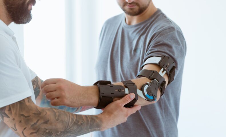
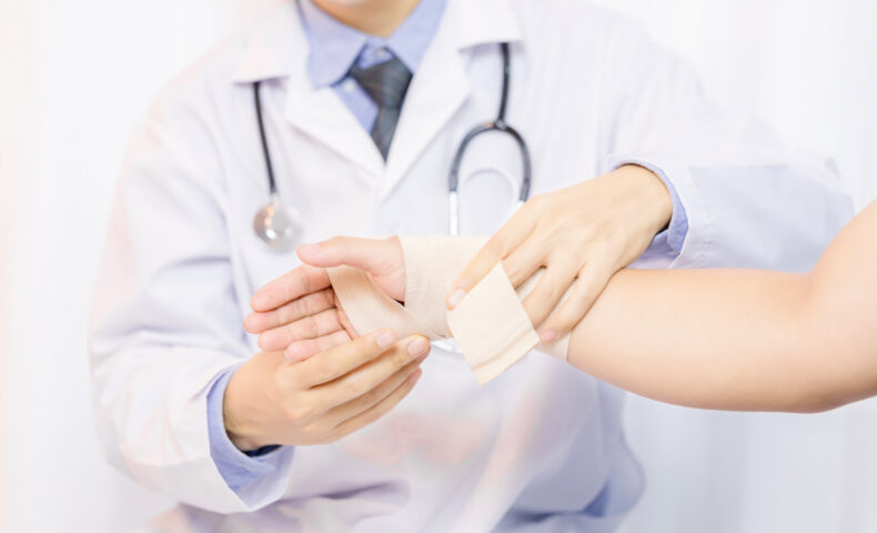
- Designing custom orthoses (elbow splints, wrist orthoses, humero-radial orthoses, etc.)
- Creating transhumeral or partial arm prostheses
- Recommending compression sleeves and garments for the arm
- Monitoring the progression of arm conditions such as edema, muscle atrophy, or trauma
Unlike traditional methods such as tape measurement or plaster casting, mobile 3D scanning is non-invasive, fast, and precise. Moreover, it integrates seamlessly with orthopedic CAD software, streamlining the entire digital design workflow.
How does a mobile 3D arm scan work?
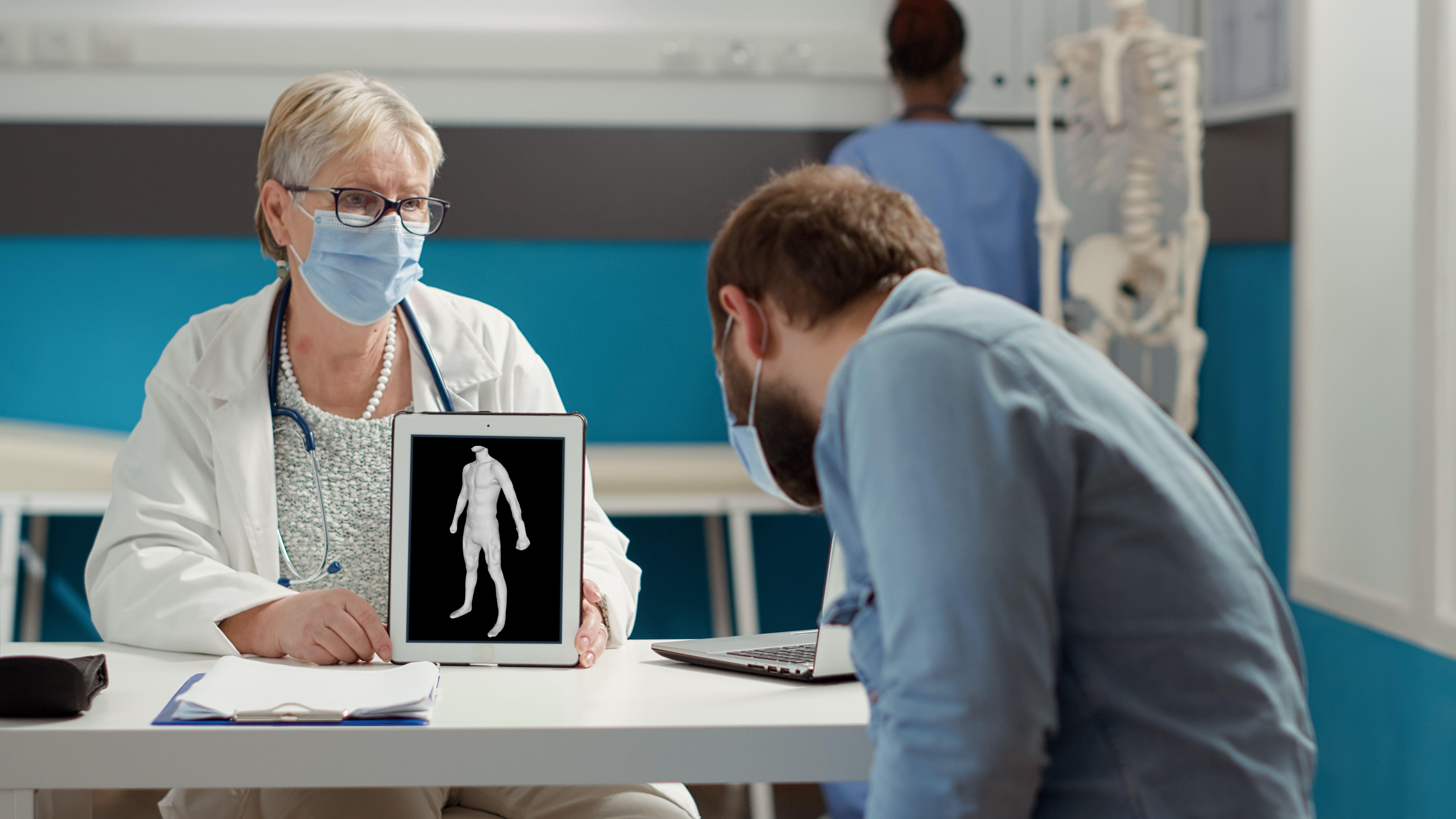
Thanks to photogrammetry, an iOS or Android smartphone can be in a 3D arm scanner. At MyFit Solutions, this technology powers our mobile 3D scanner designed for upper-limb orthopedics. Indeed, our 3D scanning experience are developed to be fast an easy to use for healthcare professionals. Here are the 4 essential steps to perform a 3D scan of the arm:
Step 1: Download a mobile 3D scanning app
Start by downloading a 3D body scanning app compatible with iOS or Android. Our app, My3D Scanner, allows precise 3D arm scanning—from the wrist and elbow to the full upper limb—for both orthopedic and therapeutic use.
Step 2: Prepare the scanning environment
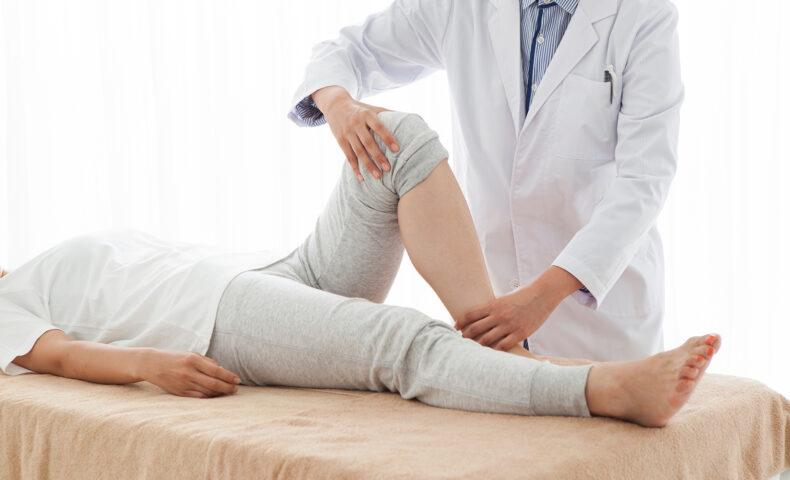
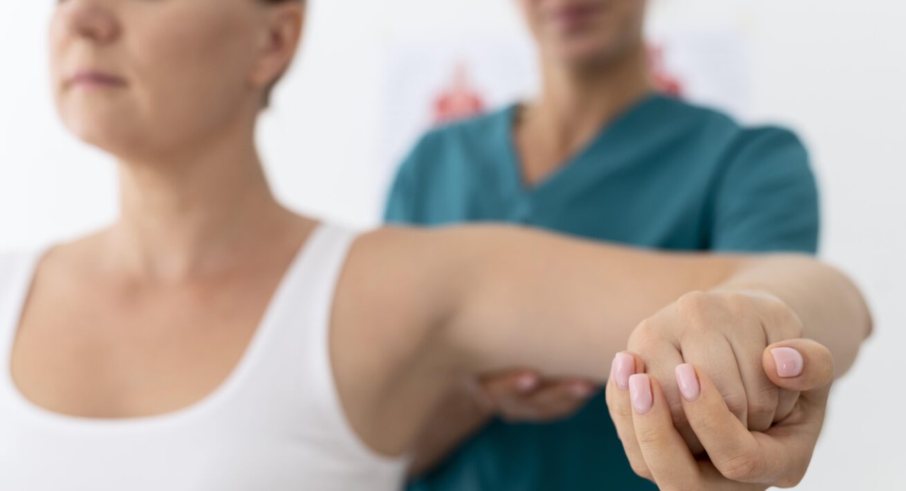
Ensure uniform lighting without shadows. Avoid reflective surfaces and keep the right scanning distance. The patient should remain still, with the arm relaxed or in the appropriate position: at the side, raised, or bent (e.g. when scanning the elbow joint).
Step 3: Capture the arm in 3D
Launch the app and follow the instructions. The scan is performed using the rear camera, which captures a series of images around the arm. These are processed through photogrammetry to create a high-precision 3D model of the upper limb.
Step 4: Export and use the 3D model
The 3D model is available instantly in the app or via our secure web platform. It can be exported in .stl or .obj format and integrated directly into orthopedic CAD tools, or used for 3D printing custom orthoses or prostheses.
Applications of mobile 3D arm scanning in orthopedics
Custom orthoses and arm prostheses
Firstly, mobile 3D arm scanning captures the complete morphology of the upper limb. A 3D model with millimetric accuracy essential for designing custom medical devices.
- Orthopedic arm orthoses: Whether for the elbow, wrist, hand, or full arm, 3D scanning enables the creation of perfectly fitted support, correction, or stabilization orthoses.
- Arm prostheses: For amputees, the 3D arm scan provides essential data for designing personalized transhumeral or partial prosthetic limbs.
- Postoperative orthoses: After surgery or trauma, clinicians can rapidly design immobilization or recovery aids based on a precise 3D model of the affected arm.
This approach empowers orthotists and prosthetists to offer personalized solutions, reduce post-manufacturing adjustments, and improve patient comfort and mobility.
Recommending compression sleeves and garments
Then, 3D scanning from mobiles delivers accurate, repeatable measurements to help recommend the best-fitting compression devices.
- Compression sleeves for lymphedema: High-precision data ensures an optimal fit, essential for effective lymphatic drainage, pain reduction, and improved quality of life.
- Post-surgical compression garments: After surgery or mastectomy, custom-fitted sleeves and garments help reduce swelling and support healing with the added benefit of non-invasive measurement.
- Medical compression wear: For burn recovery, aesthetic treatments, or dermatological conditions, the anatomical accuracy of 3D scanning ensures optimal fit and comfort.
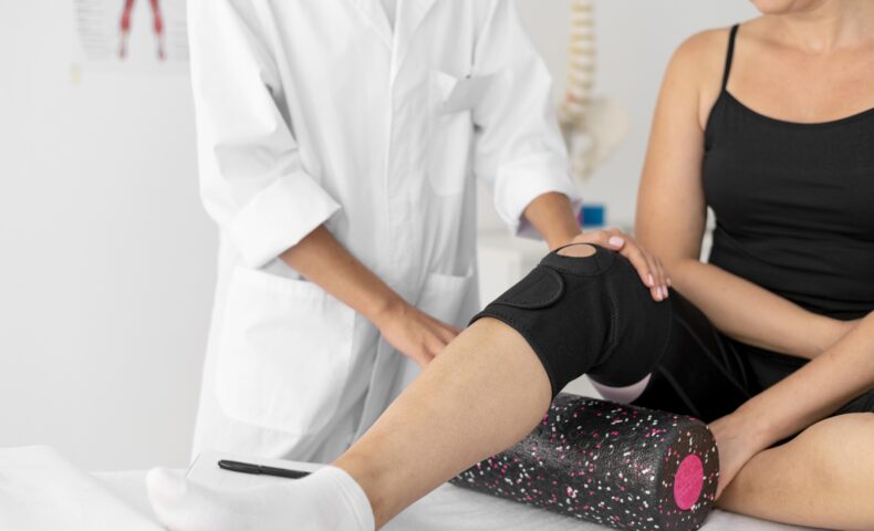
Tracking arm morphology during rehabilitation
Finally, the 3D arm scan is also a powerful tool for monitoring upper-limb rehabilitation.
- Post-traumatic edema monitoring: Track changes in swelling to adjust treatment, such as compression garments, accordingly.
- Muscle atrophy and recovery tracking: Detect and monitor asymmetry, volume loss, or functional recovery using objective data.
- Anatomical data archiving: With regular 3D scans, healthcare providers can store long-term patient data to support continuous care and treatment evolution.
In a nutshell, thanks to its ease of use, digital compatibility, and smartphone accessibility, mobile 3D scanning integrates effortlessly into clinical practice for fast, accurate, and fully digital care of the upper limb.