In short
Photogrammetry carries a mixed reputation among orthotics professionals who first encountered it through older systems — bulky, far from mobile, and reliant on a complex scaling process. MyFit mobile photogrammetry builds on the same fundamentals but adds a proprietary algorithm that stays resilient across variable lighting conditions, along with MyFit’s own scaling process — usable in a clinic, at home, or in store. The result: a photorealistic, scale-accurate 3D model from smartphones, compatible with medical use, with no specialized hardware or accessory required.
What is photogrammetry?
Photogrammetry is a 3D modeling technique based on photographic shots to determine the shape, dimensions, and position of an object in space. By using images taken from different angles, it geometrically recreates an object in three dimensions.
The method involves assembling successive images by identifying matching points between pixels, in order to reconstruct a model faithful to reality.
What are the types of photogrammetry?
- Analog photogrammetry: manual measurements from film photographs, now obsolete for professional use.
- Analytical photogrammetry: uses laser scanners and image-processing software to generate precise data.
- Digital photogrammetry: relies on advanced computing techniques to process digital images. It’s the method used in mobile 3D scanning, including in medical and O&P contexts.
Why does photogrammetry have a mixed reputation among professionals?
Photogrammetry isn’t a new technology: orthotics and measurement professionals have known about it for a long time, often through older systems that left a lasting mark on its reputation.
These early setups typically took the form of closed booths or fixed rigs, equipped with dozens of cameras arranged in a 360° circle around the subject — a principle close to a photo booth, but on a much larger scale. These installations required tightly controlled lighting conditions, a significant amount of dedicated space, and were anything but mobile: impossible to move into a clinic, a patient’s home, or the field.
Beyond the sheer size of the equipment, these older systems had two major technical limitations for medical use: the scaling of the resulting 3D model remained an additional, complex, and sometimes approximate step, and the final rendering often lacked realism (texture, color, surface fidelity). These shortcomings contributed to a lasting distrust of photogrammetry among many custom medical device professionals — a reputation that no longer reflects where the technology stands today.
What MyFit mobile photogrammetry changes
What MyFit calls MyFit mobile photogrammetry relies on the same fundamentals as traditional photogrammetry, but addresses the historical limitations that shaped its poor reputation.
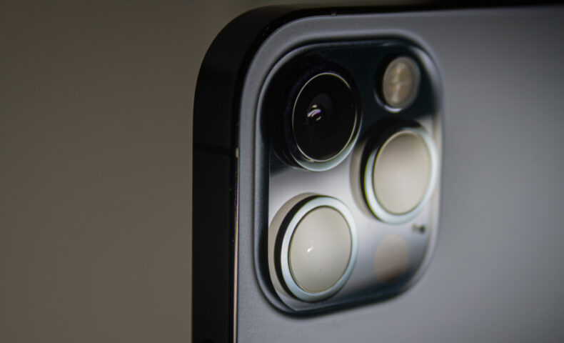
The first change is mobility. Unlike the fixed booths of the past, capture is done with a smartphone’s rear camera, with no dedicated setup. MyFit’s proprietary algorithm was designed to stay resilient across variable lighting conditions — a medical clinic, a patient’s home, a store — where older systems required tightly calibrated lighting.
The second change, arguably the most significant, is scaling. This is the step that turns a 3D model with the right shape but an undetermined size into a model with real, usable dimensions. MyFit has developed its own scaling process, which doesn’t require a dedicated depth sensor like LiDAR — a notable difference, for example, from other solutions that use a LiDAR sensor or printed reference markers of known size to calibrate the model’s scale. This innovative approach makes it possible to obtain a result that is both photorealistic and scale-accurate, using the standard capabilities of market smartphones.
The result: a faithful 3D model, at real-world dimensions, compatible with medical use — which is what sets MyFit mobile photogrammetry apart from the “legacy” photogrammetry many professionals still picture.
How does MyFit mobile photogrammetry work?
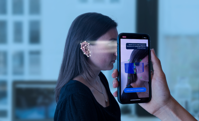
MyFit mobile photogrammetry works on a principle close to human vision: by capturing enough overlapping images from different angles, high-performance algorithms can reconstruct a three-dimensional model of the object, body part, or scanned area. To then obtain an object with the correct dimensions, a scaling process is then applied, using the phone’s standard functions
How do you perform a MyFit mobile photogrammetry scan?
Step 1: calibration
Before capture, the system initializes its sensors and its scaling process, to ensure the final 3D model reflects the real dimensions of the scanned area.
Step 2: image acquisition
A few dozen photos are generally needed to scan an area with sufficient detail. Normal lighting conditions (no major backlighting) and keeping the subject still during the shots are enough to guarantee a usable result.
Step 3: 3D modeling
The images are then processed by powerful algorithms, combined with AI, to produce a 3D mesh a structure made of many small triangles defining the object’s surface.
Step 4: post-processing
The generated model is then cleaned up: surface smoothing, removal of the background or unwanted elements, scaling, or calculation of the associated digital measurements.
What’s the difference between a 3D scanner and MyFit mobile photogrammetry?
A dedicated 3D scanner (LiDAR, Structure Sensor) generates a point cloud from a beam of light, generally with high precision but specific hardware required. MyFit mobile photogrammetry relies on a series of photos taken from different angles using the standard capabilities of market smartphones, with no need for dedicated sensors or accessories — a relevant approach where access to a specialized scanner is limited, impractical, and costly, for example in a multi-site deployment.
What are the benefits of MyFit mobile photogrammetry for professional use?
- No specialized hardware: capture is done with a smartphone’s standard rear camera, with no depth sensor or accessory to buy.
- Cross-platform compatibility: unlike TrueDepth (limited to Apple devices with Face ID) or the Structure Sensor (an iPad accessory), MyFit mobile photogrammetry works across a wide range of iOS and Android devices.
- Color and texture rendering: the generated model retains the colors and textures of the scanned area, useful for visualization and analysis.
- Proprietary scaling: a dedicated process delivers a model at real-world dimensions, with no additional depth sensor.
- Scalability: the absence of dedicated hardware makes it easier to deploy across multiple users, sites, or teams.
LiDAR, TrueDepth, Structure Sensor, MyFit mobile photogrammetry: how do they compare?
No discussion of professional 3D scanning is complete without mentioning the Structure Sensor, a long-standing reference in the industry. Like TrueDepth, it relies on infrared-assisted stereoscopy, but it’s a distinct sensor: an independent hardware accessory, developed by Structure, that attaches to an iPad.
| Criteria | LiDAR (iPhone/iPad Pro) | TrueDepth (iPhone and iPad with Face ID) | Structure Sensor (iPad accessory) | MyFit mobile photogrammetry (iOS and Android) |
| Principle | Time of flight (ToF) | Infrared-assisted stereoscopy | Infrared-assisted stereoscopy | Image processing via algorithms |
| Sensor type | Built into the device (Pro models only) | Built into the device (front camera) | Dedicated accessory, independent of the smartphone | No dedicated sensor: standard rear camera |
| Compatibility | iPhone Pro / iPad Pro | iPhone and iPad with Face ID | iPad | iOS and Android smartphones and tablets |
| Required accessory | None | Mirror, for professional use | Sensor attached via a bracket | None |
| Resolution | Low (~256×192 px) | Medium (~640×480 px) | High (up to 1040×1200 px) | Medium to high (configurable) |
| Applications | Large spaces, bulky objects | Human body, clinical use | Human body, clinical use | Human body, clinical use |
To go further, see our dedicated articles on LiDAR scanning and TrueDepth scanning.
What are the professional applications?
MyFit mobile photogrammetry has applications across several orthotics and medical contexts:
- Scanning limbs and the torso: 3D scanning captures the morphology of the scanned limb for designing custom orthoses and prostheses (for example, orthopedic insoles, a spinal brace, or a wrist orthosis).
- Compression garments: capturing volumes and circumferences helps fit a garment to the patient’s actual morphology, or manufacture fully custom garments for certain conditions.
- Head and face: useful for morphological analysis or designing adapted devices (glasses, helmets).
- Morphology tracking: 3D capture makes it possible to perform longitudinal monitoring, for example of the torso for scoliosis.
FAQ on MyFit mobile photogrammetry
The first professional photogrammetry systems relied on fixed booths equipped with dozens of cameras, with imprecise scaling and unrealistic rendering. MyFit mobile photogrammetry is built on a proprietary algorithm and scaling process that address these historical limitations.
It provides 3D models and digital measurements that are accurate for designing custom devices and for morphology tracking.
LiDAR uses a dedicated sensor to measure distance via time of flight. MyFit mobile photogrammetry reconstructs a 3D model from multiple photos, with no depth sensor, which makes it compatible with a wider range of iOS and Android devices.
Designing orthoses and orthopedic insoles, compression garments, size recommendation, morphology tracking, and more broadly any need for 3D scanning and digital measurement.