Scoliosis monitoring is essential to support patients, especially during growth. It helps observe how the back changes over time, adapt care and better document morphological changes.
With mobile 3D scanning, it becomes possible to capture the torso from a smartphone. This approach can help healthcare professionals obtain complementary morphology data, useful for screening, assessment or patient monitoring.
In this article, discover how MyFit Solutions supports NSite Medical in the development of an innovative solution. It combines 3D torso scanning, smartphone capture and artificial intelligence to contribute to scoliosis monitoring in a non-invasive and more accessible way.
From scoliosis screening to patient monitoring
Understanding scoliosis before monitoring its evolution
What is scoliosis?
Scoliosis is a deformity of the spine. It is characterized by an abnormal curvature of the spine, which can affect back alignment and trunk balance.
This deformity can affect different areas of the spine. It may have an impact on posture, the rib cage, the pelvis or certain paravertebral areas.
In some patients, scoliosis can lead to visible asymmetry of the back or trunk. It may also be associated with pain, lower back pain or a rib hump.
Scoliosis monitoring makes it possible to observe how the condition evolves over time. It helps healthcare professionals adapt care according to the patient’s age, growth and changes in body morphology.
What are the different types of scoliosis?
There are several types of scoliosis. Each form can have a different origin, progression and care pathway.
The most common forms include:
- idiopathic scoliosis: this is the most common form. It often appears during childhood or adolescence, with no clearly identified cause;
- neuromuscular scoliosis: this is linked to a muscular or neurological condition. It may be associated with conditions such as cerebral palsy or neuromuscular diseases;
- congenital scoliosis: this is present from birth. It is linked to an abnormal development of the vertebrae;
- degenerative scoliosis: this mainly affects adults. It can appear with age, in relation to disc or joint degeneration in the spine.
Identifying the type of scoliosis is important to adapt care. It is also essential to organize regular scoliosis monitoring, especially when the curvature may evolve over time.
Detecting scoliosis and organizing patient monitoring
Which examination can detect scoliosis?
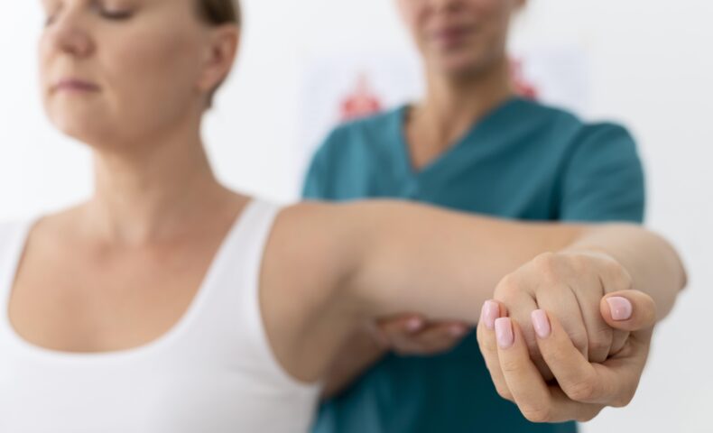
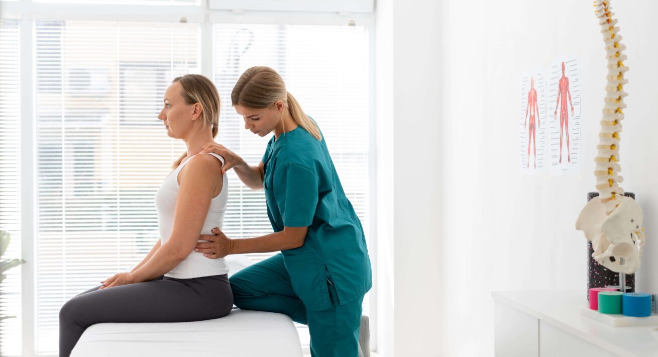
Scoliosis is generally assessed by a healthcare professional. A clinical examination can help observe posture, back alignment, trunk asymmetry or the presence of a rib hump.
X-ray remains the reference examination to confirm the diagnosis. It is used to measure the Cobb angle, which helps assess the severity of the curvature and guide care.
In children and adolescents, monitoring must be regular. Scoliosis can evolve during growth. However, repeated X-rays involve exposure to ionizing radiation, which can be a limitation in scoliosis monitoring.
This is where non-invasive solutions can provide useful complementary information. A 3D torso scan does not replace X-ray or medical advice, but it can help observe morphological changes in the back between two examinations.
Who should you consult for scoliosis?
Scoliosis may be identified by a general practitioner, school doctor, sports doctor or physiotherapist. Patient monitoring can then involve several healthcare professionals, depending on the patient’s age and the evolution of the curvature.
In case of doubt, it is important to consult a qualified professional. They can recommend the appropriate examinations and propose a consistent care pathway.
Treating scoliosis: what care options are available?
Scoliosis care depends on several factors: the type of scoliosis, its level of progression, the patient’s age and the risk of further evolution.
Depending on the situation, several approaches may be considered:
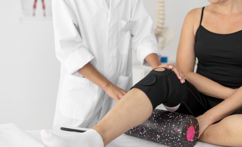
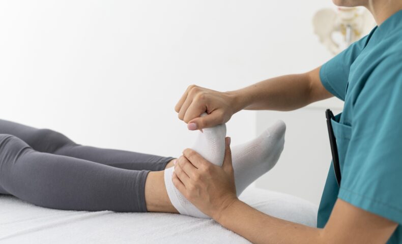
- physiotherapy: it can help strengthen the back muscles, improve posture and support mild to moderate scoliosis;
- orthopedic insoles: they may be recommended when posture is influenced by an imbalance in the lower limbs;
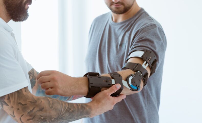
- medical brace: it may be prescribed for certain progressive forms of scoliosis, especially in growing adolescents;
- surgery: it is usually reserved for severe cases or situations where other treatments are not sufficient.
The goal is not always to fully “correct” scoliosis. In many cases, the priority is to limit its progression, preserve mobility and improve the patient’s quality of life.
Regular scoliosis monitoring therefore helps adapt care over time. It allows healthcare professionals to observe back changes and adjust treatments according to the patient’s needs.
NSite Medical x MyFit Solutions: scoliosis monitoring via smartphone
Who is NSite Medical?
NSite Medical is a US-based company from Stanford University. It develops solutions dedicated to scoliosis assessment and monitoring.
With the National Scoliosis Clinic, NSite Medical is working on an innovative approach. The goal is to provide a non-invasive, radiation-free tool that can support healthcare professionals in patient monitoring.
This technology combines artificial intelligence algorithms with MyFit Solutions’ 3D torso scanning. It could help observe morphological changes in the back, either in clinic or remotely.
The challenge is important: making scoliosis monitoring more accessible, more regular and less restrictive for patients, especially during growth.
NSite Medical x MyFit Solutions: obtaining a 3D torso scan
Who is NSite Medical?
NSite Medical is a US-based company from Stanford University. It develops solutions dedicated to scoliosis assessment and monitoring.
With the National Scoliosis Clinic, NSite Medical is working on an innovative approach. The goal is to provide a non-invasive, radiation-free tool that can support healthcare professionals in patient monitoring.
This technology combines artificial intelligence algorithms with MyFit Solutions’ 3D torso scanning. It could help observe morphological changes in the back, either in clinic or remotely.
The challenge is important: making scoliosis monitoring more accessible, more regular and less restrictive for patients, especially during growth.
Obtaining a 3D torso scan to monitor back changes
A 3D torso scan makes it possible to observe several elements related to back morphology. It can help visualize trunk asymmetry, postural alignment or visible changes over time.
The main benefit is the ability to compare captures of the same patient at different moments. Healthcare professionals therefore have a visual and digital basis to document back changes between two medical examinations.
This data does not replace reference measurements, such as the Cobb angle obtained by X-ray. However, it can enrich scoliosis monitoring with a non-invasive, regular and more accessible approach.
How to monitor scoliosis with a smartphone
With mobile 3D scanning, it is possible to capture a patient’s torso from a smartphone. The scan is guided step by step to help the healthcare professional obtain a usable capture.
This approach can provide useful complementary information for scoliosis monitoring. It helps observe morphological changes in the back, document trunk asymmetry and compare data over time.
The MyFit Solutions 3D scanning solution offers several benefits for healthcare professionals.
Accessibility
The scan can be performed in clinic, in practice or remotely.
Simplicity
The capture is performed from a smartphone, without heavy equipment.
Patient monitoring
3D data can help observe how the back changes over time.
Non-invasive approach
The 3D torso scan does not rely on X-rays.
Complementary data
3D models can enrich assessment and monitoring.
Mobile 3D scanning does not replace medical diagnosis or reference examinations. However, it can help professionals monitor morphological changes more regularly in patients with scoliosis.